
|
RECENT ENTRIES |
- • Flooding in Britain - 02.14
- • Sochi 2014 Olympics: Reaching the podium - 02.13
- • The 2014 Westminster Dog Show - 02.10
- • 2014 Winter Olympics Opening Ceremony in Soch - 02.07

Translate into:
(Hint: Use 'j' and 'k' keys to move up and down)
| October 20, 2010 |
Small Worlds
The Nikon International Small World Photomicrography Competition recently announced its list of winners for 2010. The competition began in 1974 as a means to recognize and applaud the efforts of those involved with photography through the light microscope. Peering into the small worlds of animal, plants and minerals using many techniques and different instruments, this year's entries brought us images of crystalline formations, fluorescent body parts, cellular structures and more, valuable for both their beauty and insight. The lovely folks at Nikon were kind enough to share some of their images here with us, be sure to click the link above to see all the winners. (29 photos total)
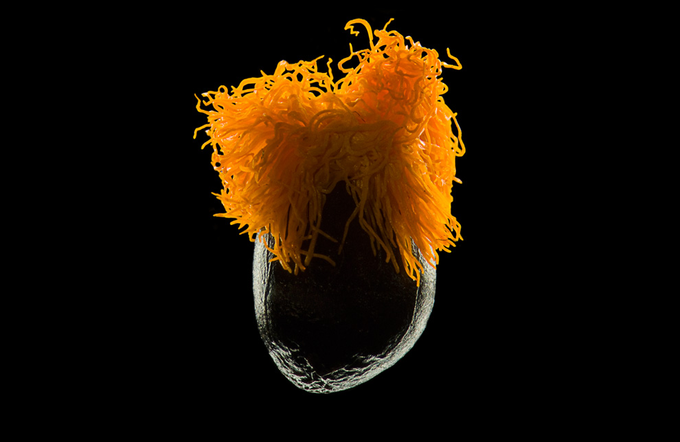
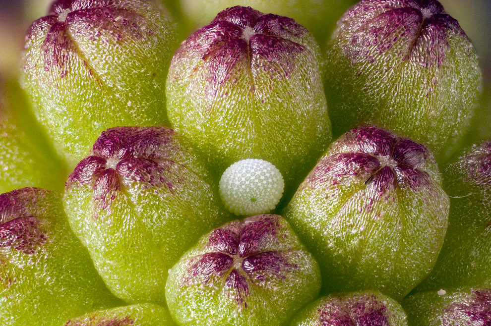
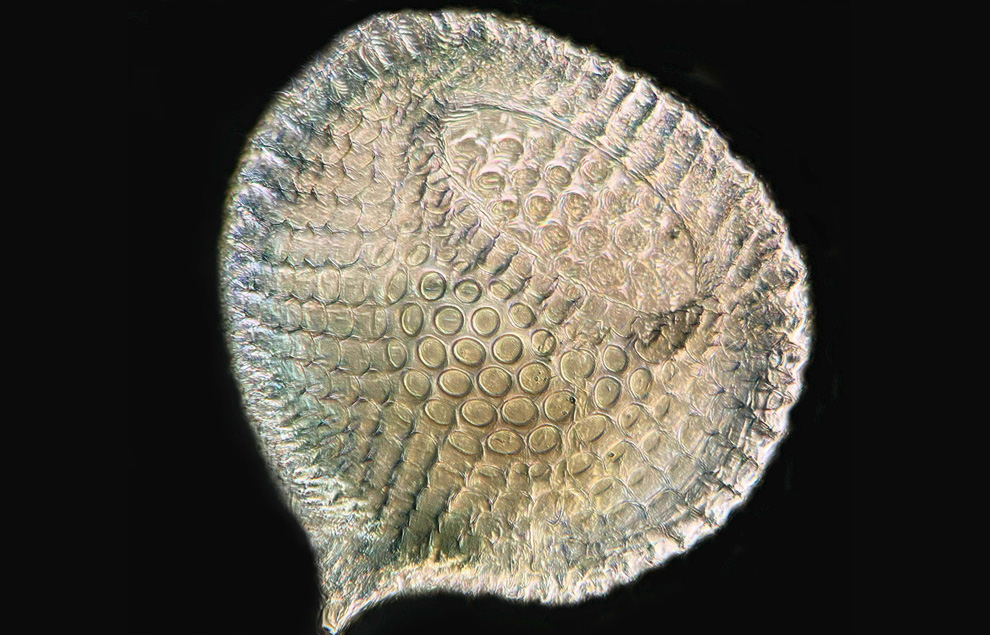
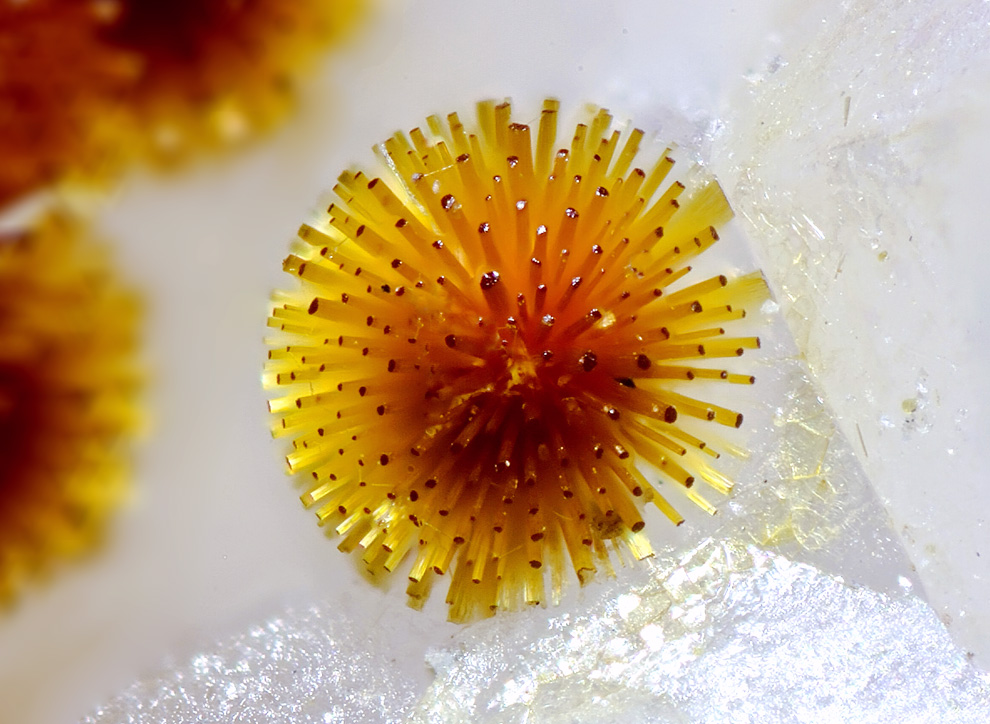
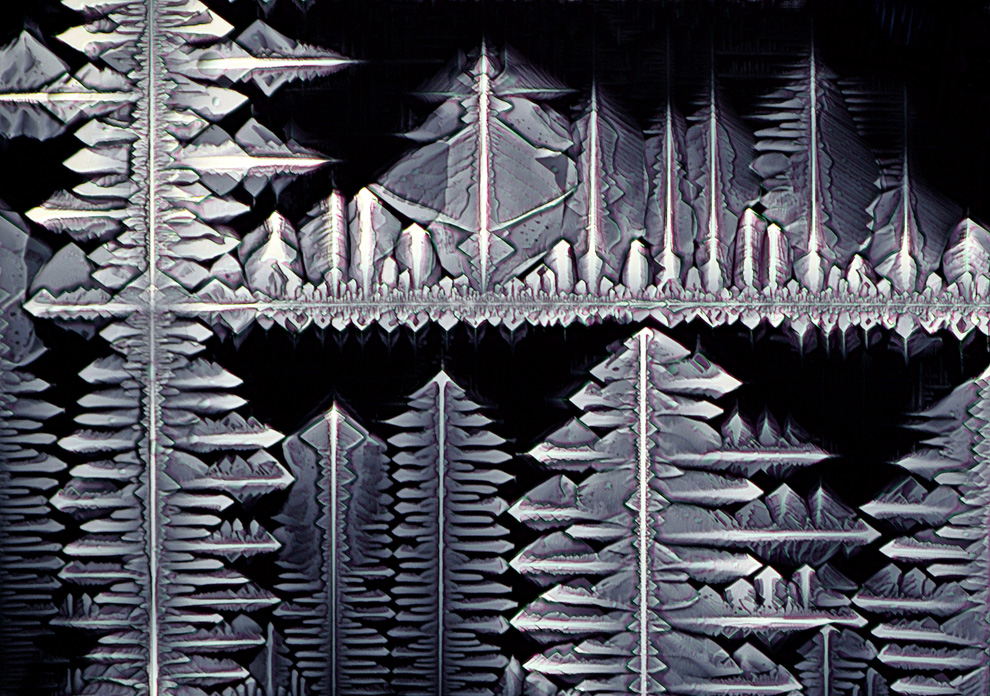
This 5th Place image of a Strelitzia reginae (bird of paradise) seed magnified 10 times comes from Viktor Sykora of the Institute of Pathophysiology, First Medical Faculty, Charles University in Prague, Czech Republic. This image was made with a stereomicroscopy technique called darkfield illumination. (Courtesy of Nikon Small World) #
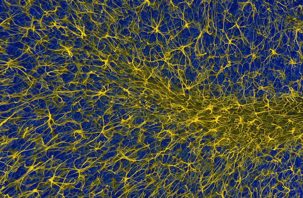
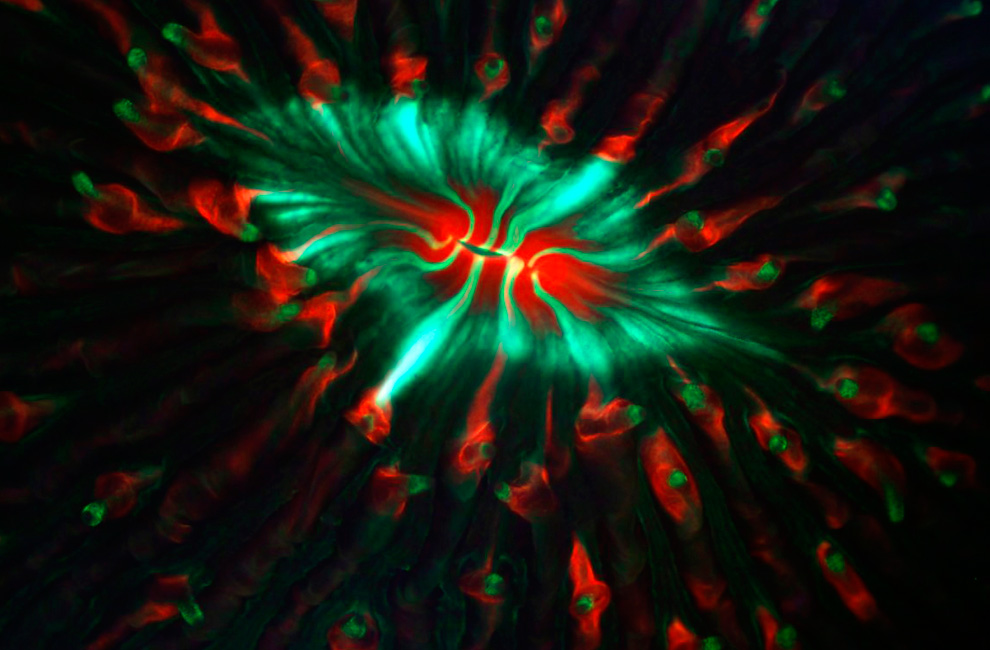
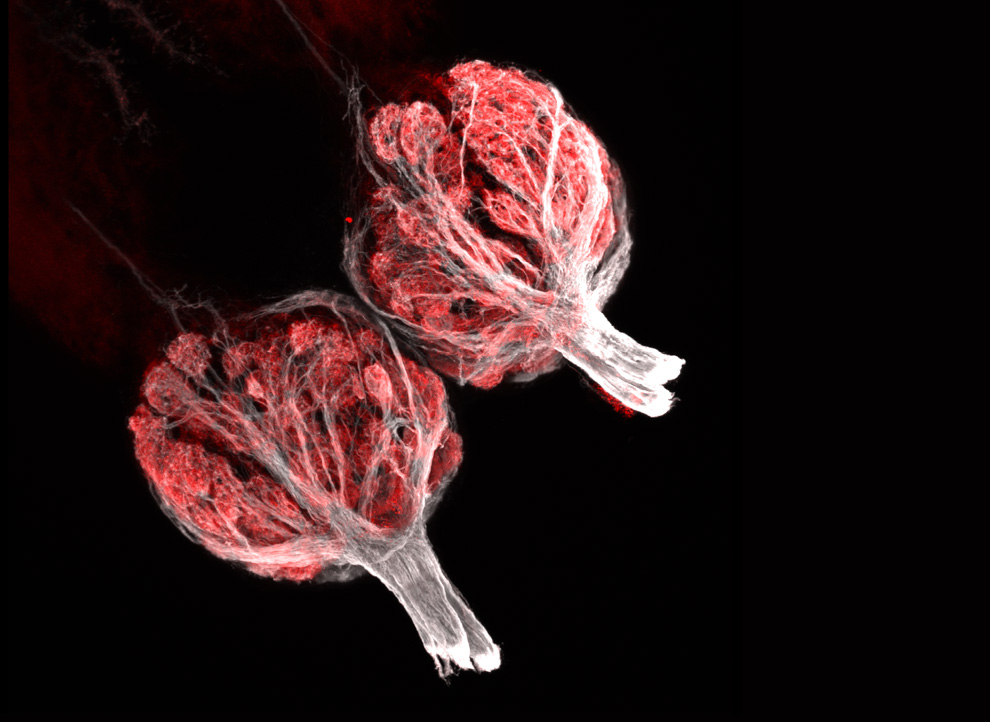
Magnified 400 times, this is a 2-Photon fluorescence image of glial cells in the cerebellum. Glial cells provide support for the brain's neurons. This image was made by Thomas Deerinck of the National Center for Microscopy and Imaging Research, University of California, San Diego. (Courtesy of Nikon Small World) #
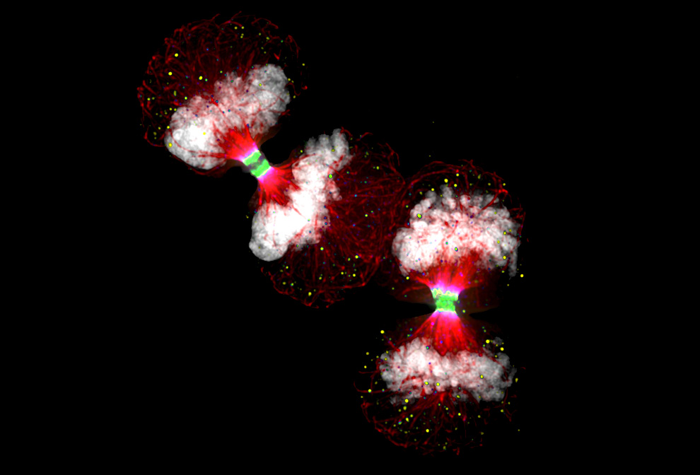
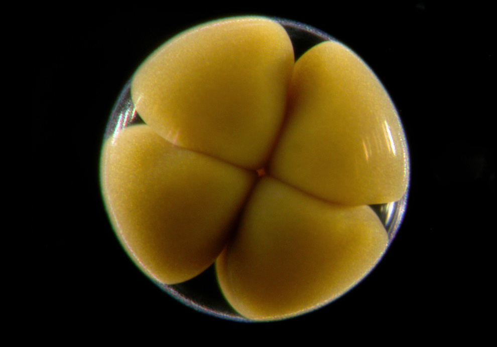
Two human cancer cells seen just before they divide into four cells, viewed at 100x magnification. This image of Telophase HeLa (cancer) cells expressing Aurora B-EGFP took 11th place and was made by Dr. Paul D. Andrews of the University of Dundee in Dundee, Scotland. (Courtesy of Nikon Small World) #
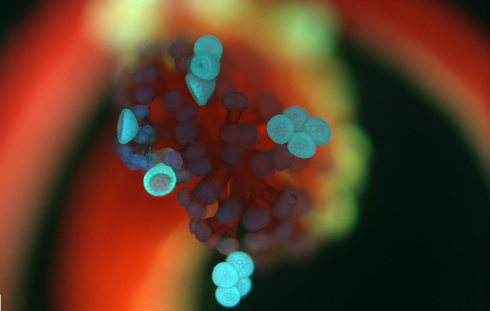
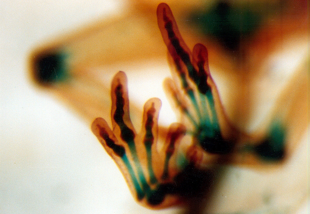
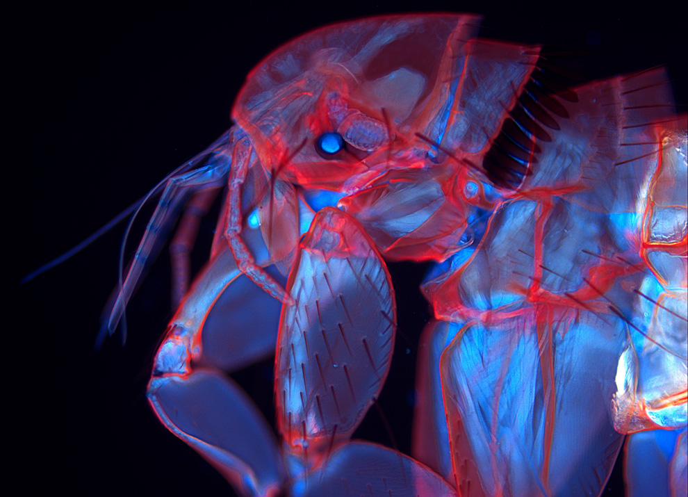
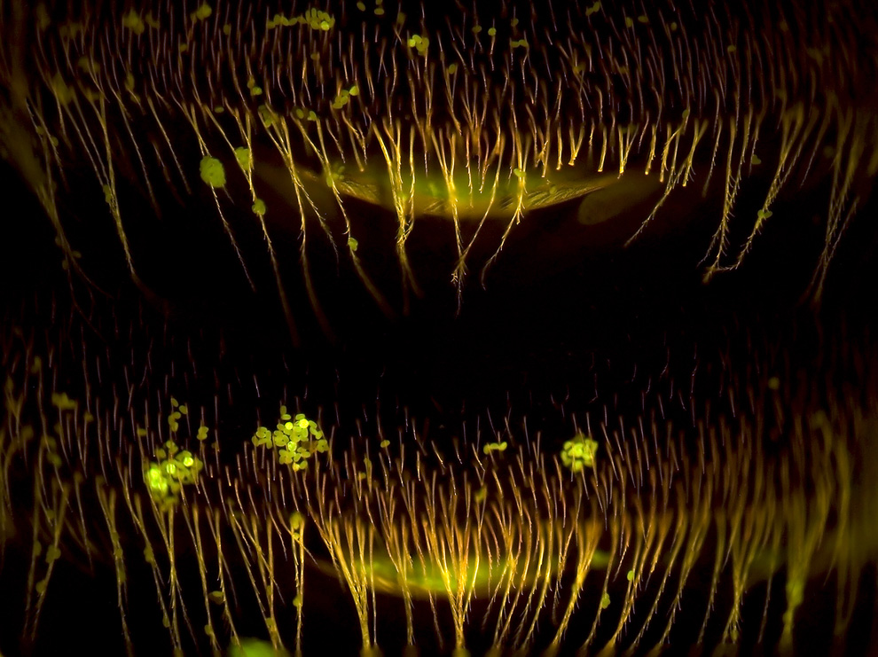
Magnified 100 times, a Mirabilis jalapa (four o'clock flower) stigma with pollen attached is seen. This 16th place image was made with epifluorescence and 3D reconstruction by Dr. Robert Markus Institute of Genetics, Biological Research Center of the Hungarian Academy of Sciences in Szeged, Hungary. (Courtesy of Nikon Small World) #
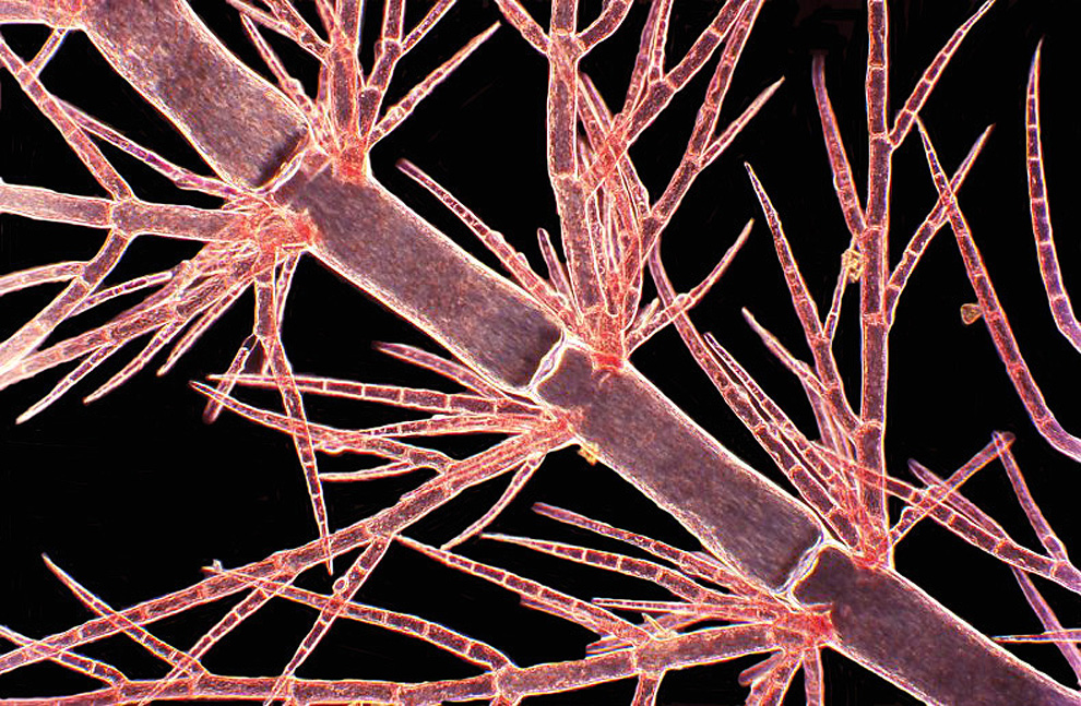
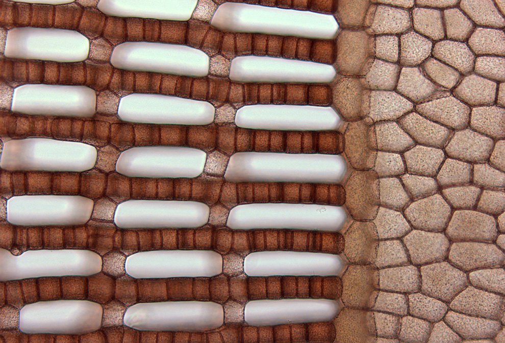
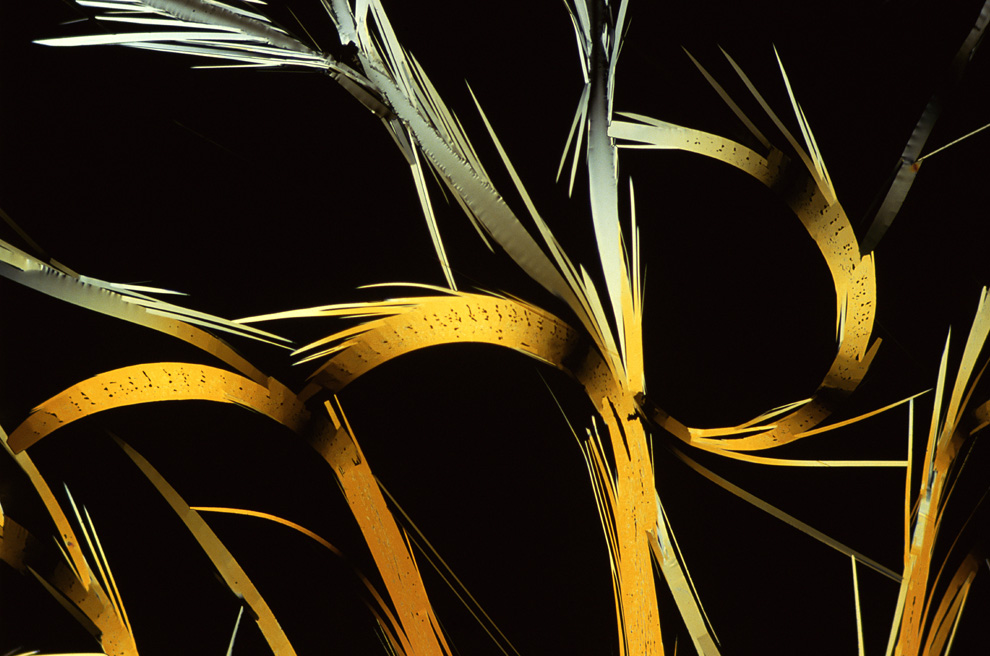
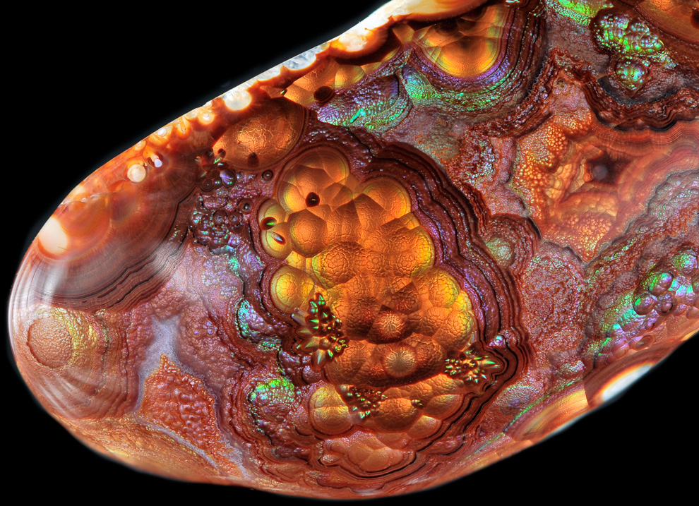
This brightfield image shows part of the structure of living specimen of Martensia sp. (red seaweed), viewed at 40x magnification. This 6th Place image was made by Dr. John Huisman of Murdoch University, School of Biological Sciences and Biotechnology in Murdoch, Australia. (Courtesy of Nikon Small World) #
More links and information
Nikon Small World - official site
Nikon Small World 2010 - Gallery of all 2010 winners and honorees